MindMap Gallery Dermatology and Venereology 004 Biogenic Skin Diseases Section 1
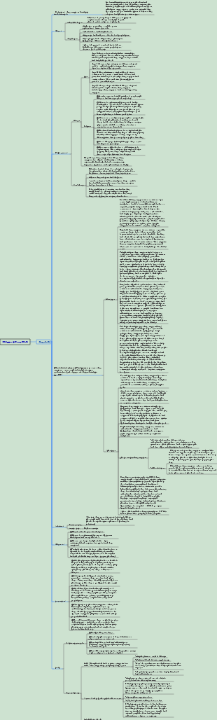
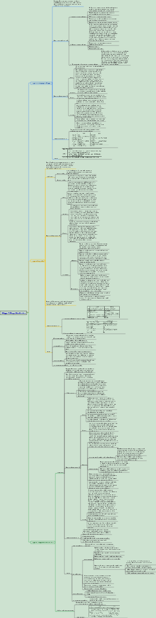
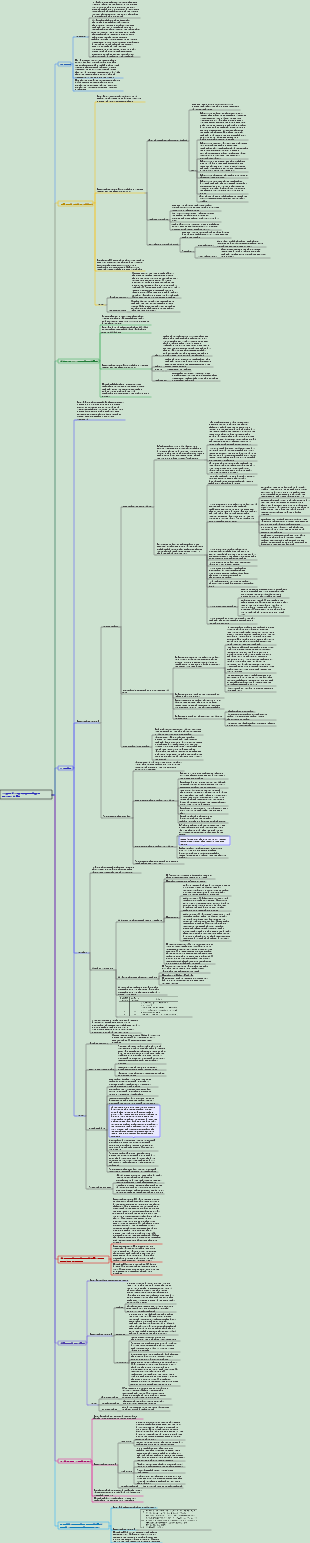
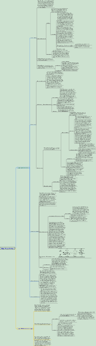
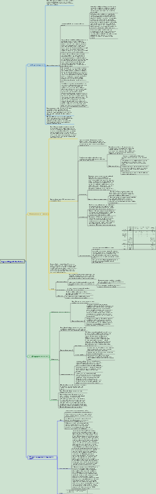
This is a mind map about Section 1 of Dermatology and Venereology 004 Biogenic Skin Diseases. Fungal skin disease is a skin infection caused by fungi. Its symptoms and diagnostic methods vary depending on the site of infection and the type of pathogen. The difference.
Edited at 2024-03-25 15:14:13Mappa mentale per l’analisi del controllo della gestione del portata di progetto. Tre sezioni principali: 1. WBS Scope Breakdown – scomposizione gerarchica del lavoro con dettaglio dello Scopo del progetto. 2. Scope Boundary / Exclusions – definizione chiara dei confini del progetto e di ciò che è escluso per evitare scope creep. Strumento ideale per project manager e team di controllo per mantenere allineamento e ridurre rischi.

Questo template, ideato con EdrawMind, è un modello completo per il miglioramento continuo del sistema di gestione della qualità, con una descrizione di circa 500 parole. È strutturato come un diagramma a lisca di pesce, strumento efficace per analizzare le cause principali dei problemi di qualità e definire le leve di controllo necessarie per risolverli. Il processo è organizzato in quattro fasi chiave: la prima è lo standard e la pianificazione, dove si definiscono gli obiettivi di qualità, i criteri di accettazione, i requisiti di prova e le porte di qualità (Quality Gate). La seconda fase analizza i processi e le porte di controllo, per garantire che ogni passaggio del lavoro segua i standard stabiliti. La terza fase riguarda l’esecuzione QA/QC, con la definizione di metodi di prova e ispezione, nonché regole per la gestione dei difetti e delle non conformità. L’ultima fase è il miglioramento e le azioni correttive e preventive (CAPA), insieme a una catena di audit per monitorare l’efficacia delle misure adottate. In basso, una tabella "Quality Gate" permette di tracciare ogni punto di controllo, con criteri specifici, metodi di prova, proprietario e stato di avanzamento. Grazie alla visualizzazione chiara e intuitiva di EdrawMind, questo strumento aiuta il team a identificare le cause root dei problemi di qualità, implementare azioni efficaci e mantenere un ciclo di miglioramento continuo, garantendo la qualità finale del prodotto o servizio.
Questo template, creato con EdrawMind, è un modello di piano di comunicazione ottimizzato, con una descrizione di circa 500 parole. È uno strumento chiave per evitare incomprensioni tra stakeholder, garantire la trasparenza e mantenere il team allineato agli obiettivi del progetto. Il modello è composto da tre elementi fondamentali e interconnessi: la matrice di comunicazione, la cadenza timeline e le regole di comunicazione/SLA. La matrice di comunicazione è una tabella dettagliata dove per ogni pubblico o stakeholder, si definisce il tipo di informazioni da condividere, lo scopo della comunicazione, il canale da utilizzare (email, riunioni, piattaforme di progetto), la frequenza, il proprietario responsabile, il formato e il percorso di escalazione in caso di problemi. La cadenza timeline è una linea temporale che definisce le scadenze delle comunicazioni chiave, garantendo che le informazioni siano condivise in momento opportuno e non si verifichino ritardi o omissioni. Le regole di comunicazione e gli accordi sul livello di servizio (SLA) definiscono le norme formali della comunicazione, come i tempi di risposta, il tono da adottare e le responsabilità di ciascun membro del team. Grazie alla struttura visuale di EdrawMind, questo template permette di pianificare la comunicazione in modo strategico, trasparente e efficiente, riducendo i rischi di cattiva informazione, migliorando la collaborazione e garantendo che tutti gli stakeholder siano informati e coinvolti nel progetto.
Mappa mentale per l’analisi del controllo della gestione del portata di progetto. Tre sezioni principali: 1. WBS Scope Breakdown – scomposizione gerarchica del lavoro con dettaglio dello Scopo del progetto. 2. Scope Boundary / Exclusions – definizione chiara dei confini del progetto e di ciò che è escluso per evitare scope creep. Strumento ideale per project manager e team di controllo per mantenere allineamento e ridurre rischi.
Questo template, ideato con EdrawMind, è un modello completo per il miglioramento continuo del sistema di gestione della qualità, con una descrizione di circa 500 parole. È strutturato come un diagramma a lisca di pesce, strumento efficace per analizzare le cause principali dei problemi di qualità e definire le leve di controllo necessarie per risolverli. Il processo è organizzato in quattro fasi chiave: la prima è lo standard e la pianificazione, dove si definiscono gli obiettivi di qualità, i criteri di accettazione, i requisiti di prova e le porte di qualità (Quality Gate). La seconda fase analizza i processi e le porte di controllo, per garantire che ogni passaggio del lavoro segua i standard stabiliti. La terza fase riguarda l’esecuzione QA/QC, con la definizione di metodi di prova e ispezione, nonché regole per la gestione dei difetti e delle non conformità. L’ultima fase è il miglioramento e le azioni correttive e preventive (CAPA), insieme a una catena di audit per monitorare l’efficacia delle misure adottate. In basso, una tabella "Quality Gate" permette di tracciare ogni punto di controllo, con criteri specifici, metodi di prova, proprietario e stato di avanzamento. Grazie alla visualizzazione chiara e intuitiva di EdrawMind, questo strumento aiuta il team a identificare le cause root dei problemi di qualità, implementare azioni efficaci e mantenere un ciclo di miglioramento continuo, garantendo la qualità finale del prodotto o servizio.
Questo template, creato con EdrawMind, è un modello di piano di comunicazione ottimizzato, con una descrizione di circa 500 parole. È uno strumento chiave per evitare incomprensioni tra stakeholder, garantire la trasparenza e mantenere il team allineato agli obiettivi del progetto. Il modello è composto da tre elementi fondamentali e interconnessi: la matrice di comunicazione, la cadenza timeline e le regole di comunicazione/SLA. La matrice di comunicazione è una tabella dettagliata dove per ogni pubblico o stakeholder, si definisce il tipo di informazioni da condividere, lo scopo della comunicazione, il canale da utilizzare (email, riunioni, piattaforme di progetto), la frequenza, il proprietario responsabile, il formato e il percorso di escalazione in caso di problemi. La cadenza timeline è una linea temporale che definisce le scadenze delle comunicazioni chiave, garantendo che le informazioni siano condivise in momento opportuno e non si verifichino ritardi o omissioni. Le regole di comunicazione e gli accordi sul livello di servizio (SLA) definiscono le norme formali della comunicazione, come i tempi di risposta, il tono da adottare e le responsabilità di ciascun membro del team. Grazie alla struttura visuale di EdrawMind, questo template permette di pianificare la comunicazione in modo strategico, trasparente e efficiente, riducendo i rischi di cattiva informazione, migliorando la collaborazione e garantendo che tutti gli stakeholder siano informati e coinvolti nel progetto.
Section 1 Fungal skin diseases
1. Dermatophytoses/tinea
【definition】
1. Ringworm: Infection of the cuticle, hair, and nail plates caused by dermatophytes is the most common infectious skin disease; it can occasionally involve deep tissues, causing fungal cellulitis, folliculitis, subcutaneous tissue abscesses, and foot ulcers. Bacteria, etc., and even hematogenous spread
2. Dermatophytids: dermatophytes and their metabolites circulate in the blood and cause allergic reactions in the skin outside the lesion; more common in summer and autumn, often occur in the acute inflammatory phase of various dermatophytosis, with dipping erosive tinea pedis Secondary bacterial infections of tinea pedis and tinea pedis are most common; clinical types include herpetic (most common), eczematoid, and erysipelas-like forms
[Cause] Dermatophytes
1. Classification
(1) According to the morphology of macroconidia: Trichophyton, Microsporum and Epidermophyton; the most common ones in my country are Trichophyton rubrum, Trichophyton gypsum and Microsporum woolly
(2) According to biological characteristics
Soil-loving: Lives in soil and is essentially soil saprotrophic bacteria; can also infect humans and animals
Animal-friendly: Lives in domestic and wild animals (cannot survive in soil), and can cause disease in humans due to contact; the number of lesions is larger but the damage is smaller, the inflammation is stronger, and the onset is more rapid.
Human-friendly: cannot survive in the soil, generally does not infect animals but only spreads among humans (including symptomatic and asymptomatic carriers); the number of lesions is small but the damage is large, the inflammation is mild, the onset is chronic and Difficult to treat
2. Biological characteristics and clinical significance
(1) Hobby for keratin: Involves the stratum corneum, hair and nail plate of the skin; the primary skin damage is superficial and the inflammation is mild
(2) Carbon and nitrogen are essential elements for growth: diabetes, long-term use of glucocorticoids, and areas with thick cuticles are susceptible to infection
(3) The optimal growth temperature is 25~28°C: it infects the surface of the body and generally does not invade deep tissues; it is prone to disease in summer and autumn
(4) Moderate humidity is required for growth and reproduction: humid, sweaty, airtight areas are susceptible to infection; ringworm is common in the south; dry environments are not conducive to growth
(5) Resistant to low temperature but not high temperature: it can be sterilized by boiling and can be stored at low temperature; skin lesions do not heal through winter, but long-term fever and bed rest can heal themselves
(6) Growth is not affected by sunlight: ultraviolet rays cannot kill
(7) The top of the hyphae grows, and the colonies grow equidistantly from the center to the surroundings: the skin lesions are ring-shaped or multi-ring-shaped, with papules and blisters on the edges, and the edges are active
(8) Colony culture grows quickly: skin lesions can appear 1 week after skin inoculation, and clinical efficacy observation generally only takes 4 weeks.
(9) Humanity, animality, and soil compatibility: different sources of infection have different clinical manifestations; the sources of infection are widespread, the incidence rate is high, and it is easy to be infected repeatedly
[Clinical manifestations, diagnosis and treatment]
(1) Tinea corporis and tinea cruris
1. Definition: Dermatophyte infection on smooth skin except the palms and soles is called tinea corporis. Tinea corporis in the external genital area of the groin and perianal area is also called tinea cruris.
2.Clinical manifestations
(1) Primary damage: Most common in summer and autumn; manifests as red papules/vesicles/papulo-herpes, the size of a pinhead/mung bean, extending equidistantly from the center to all sides, forming an annular/polycyclic shape; the edges composed of papules and blisters are narrow and Incoherent, very active, with a tendency to heal or leave temporary pigmentation in the center; significant itching
(2) Longer course of disease: ① Local infiltration and hypertrophy (chronic eczema-like changes); ② Atypical changes may occur under long-term drug use: blurred edges, reduced surface scales, loss of healing tendency in the center/edematous erythema-like appearance, which may be similar to other Skin disease that makes it difficult to identify (tinea incognito)
3. Diagnosis: Mycological examination can confirm the diagnosis
4. Treatment: ① Mainly use topical drugs, generalized tinea corporis can be treated with oral antifungal drugs at the same time; ② Maintain good personal hygiene
(2) Tinea manus and tinea pedis
1. Definition: Dermatophyte infection of the skin between the fingers (toes) and the skin of the palms and soles
2.Clinical manifestations (typing): often transform into each other
(1) Squamous blister type: the most common; it starts out as a pinhead-sized blister with thick and shiny walls, which can fuse into multi-locular bullae. When the blister wall is peeled off, a honeycomb base and a bright red eroded surface can be exposed, with varying degrees of inflammatory reactions. and itching; desquamation after drying, the scales are in the form of drops, fish scales, rings (collar-like), and continue to fall off
(2) Impregnated erosion type/intertriginous type: more common between the toes (especially the 4th and 5th toes); the skin is impregnated and white, the base is flushed, there may be exudation, and severe itching; it is prone to secondary bacterial infection (such as acute lymphangitis, cellulitis, erysipelas)
(3) Hyperkeratosis type: The skin is rough and thickened, with obvious keratinization, and is prone to chapping; it is more common in patients with a longer course of disease, and generally there is no obvious itching.
3. Treatment: ① Treatment by type: compound benzoic acid liniment and antifungal cream can be used for the scaly blister type; antifungal cream can be used for the immersion and erosion type, and bacterial infection must be controlled first; compound benzoic acid liniment and antifungal cream can be used for the hyperkeratosis type. Formic acid ointment or miconazole cream, urea grease can be added for those with chapped skin; ② Oral antifungal drugs can be used in severe cases
(3) Tinea capitis
1. Definition: Dermatophyte infection involving the scalp and hair
2. Clinical manifestations (types): divided into white tinea (the most common), black tinea, yellow tinea, and purulent tinea
Mother and child spots
Several smaller identical lesions appear around the lesion
yellow scab
It appears as a light yellow scab about the size of a soybean, with raised edges and a saucer-shaped center attached to the scalp. It can expand and fuse, and in severe cases, covers the entire scalp. After the scab is removed, there will be a flushed and eroded surface underneath.
Kerion: local allergic reaction on the scalp caused by dermatophyte infection; manifested as one/several abscesses with obvious inflammation, neat edges, and a sense of fluctuation; they can merge into plaques with clear, sharp edges as neat as a knife cut ; The hair in the skin lesions is loose and easy to break; the onset is rapid, fever, poisoning symptoms, and lymph node enlargement may occur, and scars and permanent hair loss may be left after recovery.
3. Treatment: ① Antifungal drugs: oral and topical; ② Shaving and shampooing; ③ Avoid contact with asymptomatic carriers, cats and dogs, and pay attention to disinfection; ④ Do not incision and drainage of pus ringworm (to avoid the formation of larger permanent scars) )
(4) Onychomycosis (onychomycosis)
1. Definition: Fungal infection of the nail plate and nail bed; among them, dermatophyte infection is also called tinea unguium.
2.Clinical manifestations (typing)
(1) Distal lateral subungual type (DLSO): the most common; the fungus first invades the cuticle at the distal and lateral edges of the nail plate, and then gradually spreads to the nail bed; causing thickening of the cuticle, subungual hyperplasia, and The end deck lifts, creating a gap between the nail bed and the deck
(2) White superficial type (SWO)/fungal leukonychia: There are clearly defined, opaque dots and strips of white lesions on the nail surface, which turn yellow over time and make the nail brittle and easily broken; caused by fungi on the nail surface direct vaccination
(3) Proximal subungual type (PSO): Pathogens invade from the proximal nail groove and form white spots at the root of the nail, which can grow with the nail plate until it extends to the entire nail plate; the accumulation of keratin under the nail is not obvious
(4) Total nail damage type (TDO): Developed from the above three types; it is characterized by thickening, brittleness, incompleteness, dirt, unevenness of the whole nail, or even the disappearance of the whole nail, leaving exposed cutin and thickened nail bed
3. Diagnosis: The diagnosis relies on mycological examination; the following symptoms are helpful for diagnosis: ① It is often accompanied by dermatophytosis in other parts of the body; ② For tinea manuum that can be diagnosed, the associated onychomycosis is mostly onychomycosis; ③ If there are multiple onychomycoses The nail plate is involved and the process is gradual and slow. The severity of the nail disease varies. It is often considered to be onychomycosis.
4. Treatment: ① Locally encapsulate the diseased nail with 40% urea lipid for 1 week, scrape it off with a knife after softening, and then apply 3% miconazole tincture externally until the new nail completely grows; ② Treat other ringworms; ③ Oral antifungal drugs
2. Candidiasis
[Cause] Candida: Opportunistic pathogenic bacteria (spore phase → hyphae phase), multiple endogenous infections; Candida albicans is the most common and most virulent
[Clinical manifestations]
Skin and mucous membrane type
Oral thrush: more common in infants and young children; manifested by the appearance of white pseudomembranes on the oral mucosa, pharynx, tongue, gums, etc., with clear edges and peripheral redness; a red base can be seen when the pseudomembrane is removed
Candida vulvovaginitis: Vaginal mucosa presents as thrush-like white pseudomembrane; increased leucorrhea mixed with cheese-like/bean dregs-like particles; often accompanied by vulvar redness, swelling, and severe itching
Candidal balanitis: manifests as mildly flushed patches with a dry, smooth surface; it may also manifest as superficial erosion and thin-walled pustules; in severe cases, there may be thrush-like pseudomembranes, which may even spread to the entire body. Perineum; anterior urethral inflammation may occur when urethra is involved
Candidal interdigital erosion: more common between the 3rd and 4th fingers, often related to obesity and occupation
Candida intertrigo (candida intertrigo): more common in children and obese people, manifesting as skin flushing, erosion, exudation, and scabs in fold areas (such as in front of the neck, perianal area, groin, under the breasts, and armpits). There may be scattered red papules with scaling
Papular cutaneous candidiasis: more common in obese children; manifests as mung bean-sized hemispherical dark red papules with clear edges and gray-white round thin scales, distributed on the chest, back and perineum, not fused, and itchy; can be associated with red papules. Prickly heat concurrently
Candida paronychia: Symptoms include redness, swelling and slight pain in the nail groove, but no suppuration; the nail plate is turbid, with white spots, and the surface has transverse ridges and grooves, and is uneven but still shiny and not broken.
Chronic mucocutaneous candidiasis (CMCC)
Patients often have endocrine abnormalities/low immune function. The disease usually develops at an early age and may have a familial genetic tendency.
Thrush and erosion of the corners of the mouth usually appear first, followed by erythematous and scaly rashes with keratin hyperplasia on the head, face, and extremities; then the trunk is gradually affected; paronychia/candida infection, thinning and loss of hair may occur at the ends of some fingers. Premature appearance; verrucous proliferative skin lesions may appear on the forehead and nose, covered with oyster shell-like brown scabs, sometimes as high as 2cm, which is characteristic
Candidal granuloma/deep cutaneous candidiasis: more common in immunocompromised individuals
Systematic
Manifested as gastrointestinal candidiasis, bronchopulmonary candidiasis, urinary tract candidiasis, candidal endocarditis, candidal meningitis, etc.; mainly seen in diabetes, malignant tumors, various chronic wasting diseases, and AIDS Many of these patients have long-term use of broad-spectrum antibiotics, glucocorticoids, radiotherapy, chemotherapy, major surgery, etc.
disseminated
Multiple systems in the body are involved at the same time, including candida septicemia; direct microscopy and culture of multiple specimens are positive at the same time and are of the same bacterial species; the prognosis is grim
[Diagnosis] Note: Positive direct microscopy (detection of pseudohyphae and spores) is more clinically significant than positive culture alone
[Treatment] ① Local treatment: suitable for skin and mucosal candidiasis; ② Systemic treatment: suitable for extensive skin lesions or systemic and disseminated candidiasis
[Prevention] High-risk patients can take fluconazole 50 mg or itraconazole 100 mg daily to prevent candida infection.
3. Pityriasis versicolor/tinea versicolor/sweat spots
[Cause] A chronic, asymptomatic fungal infection in the superficial part of the stratum corneum caused by Malassezia furfur; the onset is associated with high temperature and humidity, excess fat and sweat, malnutrition, chronic diseases, and the use of sugar related to corticosteroids
[Clinical manifestations] It usually occurs in areas rich in sebaceous glands. In infants and young children, the glabella, middle eyebrow, forehead, and temples are most common and manifest as hypopigmented spots; ② The primary lesion is a mung bean-sized macule without inflammation; it is covered with pityriasis-like spots. Small shiny scales, which can leave temporary hypopigmented spots after removal; the appearance of the infected area is a patchwork of dark, light and normal skin colors; ③ Generally there are no subjective symptoms, and occasionally mild itching; patients are prone to sweating , hair grows in summer and retreats in winter
[Diagnosis] Scraping off scales and direct microscopic examination can reveal tinea versicolor (appearing as "banana and grape"), which has diagnostic value
4. Pityrosporum folliculitis (pityrosporum folliculitis)
[Cause] Chronic hair follicular infection caused by Malassezia furfur
[Clinical Manifestations] Chronic onset, usually occurring on the upper back, upper chest, shoulders and forearms, more common in young people and more common in summer; typical manifestations are dark red dome follicular papules/pustules, isolated and scattered, and may gather but not merge. ; There is redness around the area, and powdery substance may be extruded; it may be consciously itchy, aggravated by sweating; it may be accompanied by seborrheic dermatitis
5. Sporotrichosis
[Definition] It is a subacute and chronic infection of the skin, subcutaneous tissue and peripheral lymphatic system caused by Sporothrix shenckii. It can occasionally involve bones and internal organs, and even cause disseminated infection.
[Clinical manifestations]
Skin type
It commonly occurs on the limbs and face; it can be divided into fixed type (the lesions are only seen at the initial sore site, and typically appear as nodules, chancre-like), and lymphatic type (the lesions are mainly nodules arranged along the lymphatic vessels, and there may be nodules. scabs and ulcers; lymphatic vessels between skin lesions often become hardened and cord-like, but local lymph nodes are generally not swollen), disseminated type
Leather appearance
Invasion of various tissues throughout the body; clinical manifestations are non-specific
[Examination] Pathology: Early manifestations are non-specific granulomas in the dermis; PAS staining shows round, cigar-shaped spores and stars.
[Treatment] Potassium iodide is the drug of choice
6. Chromoblastomycosis
[Characteristics] It is a group of chronic infections of the skin and subcutaneous tissue caused by dark fungi; there is often a history of local trauma. It is initially a single inflammatory papule at the site of fungal invasion, and gradually expands to form dark red nodules/plaques with dark surrounding areas. The red inflammatory infiltration zone may be covered with brown scabs on the surface. There are small black-brown dots about the size of a pin cap scattered on the scabs. There is often pus overflowing under the scabs. Granular/papillary granulations can be seen after uncovering them. In the late stage, they can spread along lymphatic vessels. scattered