MindMap Gallery Medical Physics-Radiotherapy Quality Control and Radiation Protection
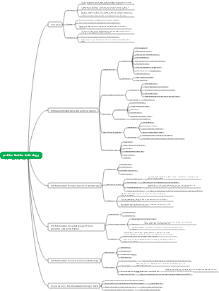
This is a mind map about medical physics, radiotherapy quality control and radiation protection. It is introduced in detail and comprehensively described. I hope it can be helpful to interested friends.
Edited at 2023-11-29 21:07:50This is a mind map about bacteria, and its main contents include: overview, morphology, types, structure, reproduction, distribution, application, and expansion. The summary is comprehensive and meticulous, suitable as review materials.

This is a mind map about plant asexual reproduction, and its main contents include: concept, spore reproduction, vegetative reproduction, tissue culture, and buds. The summary is comprehensive and meticulous, suitable as review materials.
This is a mind map about the reproductive development of animals, and its main contents include: insects, frogs, birds, sexual reproduction, and asexual reproduction. The summary is comprehensive and meticulous, suitable as review materials.
This is a mind map about bacteria, and its main contents include: overview, morphology, types, structure, reproduction, distribution, application, and expansion. The summary is comprehensive and meticulous, suitable as review materials.
This is a mind map about plant asexual reproduction, and its main contents include: concept, spore reproduction, vegetative reproduction, tissue culture, and buds. The summary is comprehensive and meticulous, suitable as review materials.
This is a mind map about the reproductive development of animals, and its main contents include: insects, frogs, birds, sexual reproduction, and asexual reproduction. The summary is comprehensive and meticulous, suitable as review materials.
Radiotherapy Quality Control and Radiation Protection
Radiotherapy quality control
Concepts of Quality Assurance and Quality Control in Radiation Therapy
quality assurance
QC
The need to perform quality assurance
Quality Assurance (QA) Purpose
Reduce uncertainties and errors in planning, instrument performance, and treatment verification, ensure treatment accuracy and equipment precision, and improve efficacy.
Radiotherapy quality assurance content
quality assessment
QC
Quality management in the radiotherapy process
Quality assurance of target delineation
After simulation and two-dimensional positioning, the field of view is outlined on the patient's body surface.
After CT positioning, the tumor target volume is delineated on the generated patient image data, including the tumor target volume (GTV) and the clinical target volume (CTV). Internal target volume (ITV), planning target volume (ptv), organ at risk (OR), and three-dimensional (3D) target volume delineation of the planning risk zone (PRV)
Characteristics of Ct simulation positioning
It provides a reference position, establishes three-dimensional spatial coordinate information, and can clearly display the patient's anatomical structure. The error variability is small, and the tissue electron density provides a basis for dose calculation.
Features of MR simulation positioning
The flat bed surface can be connected to the patient's fixture, the scanning coil can be adapted to the patient's fixture, and the inspection aperture is large (>70cm). The magnetic field is uniform, etc. In order to improve the accuracy of tumor target location, specific scanning sequences need to be used for tumors in different parts.
Characteristics of pet-ct images
It can feed back tissue metabolism signals to distinguish the level of breathability in tumor tissue and provide rich information for tumor staging.
Plan design quality assurance
Quality Assurance of Treatment Planning Systems
Treatment planning system is an acceptance test
Clinical testing of treatment planning systems
Periodic quality assurance of planning systems
The content of regular inspections mainly includes hardware, software and data transmission. According to the length of the inspection cycle, it is divided into weekly inspection, monthly inspection, quarterly inspection and annual inspection.
Quality Assurance of Patient Treatment Plans
Plan design consistency check
Independent verification of treatment plans
Machine hop count verification
Pre-treatment examination and mid-treatment weekly examination of the treatment plan
Quality assurance of treatment delivery
Review documents related to radiation therapy plan
Image-guided radiation therapy (IGRT)
Quality assurance of radiotherapy equipment
Radiotherapy equipment
Linear Accelerator
Ct simulator
Detection of laser lamp accuracy
Bed motion accuracy detection
Detection of the accuracy of the bed's advance and retreat directions
Detection of bed lifting direction
Detection of size accuracy of scanned images
Detection and air calibration of scanning CT value accuracy
The brightest parts of the image are bones, and the darkest parts are air.
After-loading treatment machine
Shandong Xinhua’s after-loading machines use Category III radioactive source Iridium-192
Quality control items
Performance of personal meters and environmental monitors, battery and backup battery power detection
Radiation protection dosimetry instruments are important to ensure operator safety
Equipment surface, ambient dose equivalent rate
The surrounding dose equivalent rate due to leakage radiation at any position 5cm away from the surface of the source is not greater than 50usv/h
On the spherical surface 100cm away from the surface of the source, the surrounding dose equivalent rate due to leakage radiation at any point is not greater than 5usv/h
For iridium-192, testing must be performed after source change or repair (WS262-2017)
Radioactive source placement accuracy
The positioning accuracy of the radioactive source is ±1mm as standard
Maximum error and cumulative error of radioactive source positioning
The maximum cumulative error standard for radioactive sources is ±2mm
Dwell time accuracy
The setting time should be similar to the average dwell time of conventional radiotherapy
Error=set time-actual measurement time, the maximum error is ±0.5s
Summarize
Full-time positioning technicians are responsible for positioning membranes for patients
Full-time physicist does equipment quality control
Specialist therapists are responsible for position verification and dose verification of patients before radiotherapy
Radioprotection
Basic knowledge of radiation protection
ionizing radiation
Radiation is the process by which electromagnetic waves or particles emit energy
Radiation source
natural radiation
sun, earth
artificial radiation
X-ray machine, nuclear weapon explosion, nuclear power plant accident
Radioactive source classification
Class I radioactive sources and hazardous sources
Without protection, exposure to such sources can kill within minutes to an hour.
Class II radioactive sources are high-risk sources
Without protection, exposure to such sources can lead to death within hours to days.
Category III radioactive sources are medium-risk sources
Without protection, exposure to such sources can cause permanent damage to humans within a few hours, and death can occur within days to weeks of exposure.
Category IV radioactive sources are low-risk sources
Basically it will not cause permanent damage to humans, but it may cause recoverable temporary damage to people who are exposed to the radioactive source at close range for a long time.
Category 5 radioactive sources are extremely low-risk sources
Will not cause permanent damage to people
Biological effects of ionizing radiation
How radiation affects the human body
External radiation exposure, including medical diagnosis and treatment
Radioactive nuclide external contamination, contaminating the surface of the human body causing localized external exposure
Internal contamination of radionuclides, inhaled into the blood through the body surface or inhaled through food, or entering the body through skin wounds to constitute internal exposure
In practice, there are often multiple irradiation methods, which simultaneously constitute composite irradiation.
Effects of Radiation on the Human Body
The effects that appear on the irradiated person after the human body is exposed to ionizing radiation are somatic effects, and the effects that appear on the descendants of the irradiated person are called genetic effects.
According to the nature of the law of effect occurrence
random effects
Deterministic effect/non-random effect
According to the time when the effect appears
recency effect
acute effects
chronic effects
forward effect
body changes
Skin damage, reproductive organ damage, hematopoietic organ damage, digestive organ damage, central nervous system damage
cataracts, compromised immune system
cancer disease
genetic disease
Biological effects of ionizing radiation
organizational sensitivity
highly sensitive organization
Lymphoid tissue, thymus, bone marrow, gastrointestinal epithelium, gonads, embryonic tissue
Moderate
Senses, skin, salivary glands, kidneys, liver, lungs
B
central nervous system, endocrine glands, heart
Not sensitive
Muscle, cartilage/bone, connective tissue
radiation damage
Total dose, dose rate and mode of exposure
occupational exposure
Exposure classification
occupational exposure
public exposure
The annual effective dose does not exceed 1 msv
The average dose for 5 consecutive years does not exceed msv
The effective dose in a given year does not exceed 5 msv
The annual equivalent dose of the eye lens is 15 msv
The annual dose for skin is 50 msv
No one under the age of 16 may receive occupational exposure
medical exposure
to staff
The average annual effective dose for 5 consecutive years does not exceed 20 msv
The effective dose does not exceed 50 msv in any year
The annual equivalent measurement of the eye lens does not exceed 150 msv
Annual equivalent dose to extremities or skin does not exceed 500 msv
For geotechnical workers or students aged 16~18
The annual effective dose does not exceed 6 msv
The single dose of adhesive for the lens of the eye does not exceed 50 msv
Annual equivalent dose to extremities or skin does not exceed 150 msv
External exposure radiation protection
protective principle
Shorten exposure time
Increase distance from radiation source
Set up protective shielding
Lead sheets, cement walls, water
Accelerator room structure
Ct protective door
accelerator protective door
There is an ionizing radiation sign outside the computer room door, and eye-catching work indicators are installed.
All computer rooms are equipped with ventilation devices
The layout of the computer room should be reasonable, and debris unrelated to diagnosis should not be piled up.
Protection against accelerator characteristics
Blocking of x-rays
Machine head structure
Multi-leaf grating blocks rays
Blocking of induced rays
Protection against ozone
Protection of staff
Act according to the three protection principles of time, distance and shielding
Do a blood routine once every six months to observe changes in white blood cells
Internal exposure radiation protection
Safety of Radioactive Sources
Patient protection lead shielding
Monitoring intercom system
Protection of staff