MindMap Gallery The Relationship between Cell Biology and Chemistry and Other Disciplines
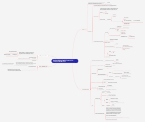
The Relationship between Cell Biology and Chemistry and Other Disciplines
The field of cell biology and chemistry explores the chemical processes within cells, such as metabolism and signaling pathways, and their relation to cellular functions. It intersects with disciplines like genetics, molecular biology, and biochemistry, providing a holistic understanding of biological systems. Biochemistry, as a bridge between biology and chemistry, focuses on the chemical reactions and pathways that occur within living organisms.
Edited at 2024-12-13 02:31:16- Recommended to you
- Outline