MindMap Gallery epithelial tissue
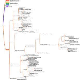
Histology and Embryology, introduces general characteristics, Covering epithelium, the special differentiated structure of epithelial tissue, etc. Hope this mind map helps you!
Edited at 2024-02-08 17:07:34Mappa mentale per l’analisi del controllo della gestione del portata di progetto. Tre sezioni principali: 1. WBS Scope Breakdown – scomposizione gerarchica del lavoro con dettaglio dello Scopo del progetto. 2. Scope Boundary / Exclusions – definizione chiara dei confini del progetto e di ciò che è escluso per evitare scope creep. Strumento ideale per project manager e team di controllo per mantenere allineamento e ridurre rischi.

Questo template, ideato con EdrawMind, è un modello completo per il miglioramento continuo del sistema di gestione della qualità, con una descrizione di circa 500 parole. È strutturato come un diagramma a lisca di pesce, strumento efficace per analizzare le cause principali dei problemi di qualità e definire le leve di controllo necessarie per risolverli. Il processo è organizzato in quattro fasi chiave: la prima è lo standard e la pianificazione, dove si definiscono gli obiettivi di qualità, i criteri di accettazione, i requisiti di prova e le porte di qualità (Quality Gate). La seconda fase analizza i processi e le porte di controllo, per garantire che ogni passaggio del lavoro segua i standard stabiliti. La terza fase riguarda l’esecuzione QA/QC, con la definizione di metodi di prova e ispezione, nonché regole per la gestione dei difetti e delle non conformità. L’ultima fase è il miglioramento e le azioni correttive e preventive (CAPA), insieme a una catena di audit per monitorare l’efficacia delle misure adottate. In basso, una tabella "Quality Gate" permette di tracciare ogni punto di controllo, con criteri specifici, metodi di prova, proprietario e stato di avanzamento. Grazie alla visualizzazione chiara e intuitiva di EdrawMind, questo strumento aiuta il team a identificare le cause root dei problemi di qualità, implementare azioni efficaci e mantenere un ciclo di miglioramento continuo, garantendo la qualità finale del prodotto o servizio.
Questo template, creato con EdrawMind, è un modello di piano di comunicazione ottimizzato, con una descrizione di circa 500 parole. È uno strumento chiave per evitare incomprensioni tra stakeholder, garantire la trasparenza e mantenere il team allineato agli obiettivi del progetto. Il modello è composto da tre elementi fondamentali e interconnessi: la matrice di comunicazione, la cadenza timeline e le regole di comunicazione/SLA. La matrice di comunicazione è una tabella dettagliata dove per ogni pubblico o stakeholder, si definisce il tipo di informazioni da condividere, lo scopo della comunicazione, il canale da utilizzare (email, riunioni, piattaforme di progetto), la frequenza, il proprietario responsabile, il formato e il percorso di escalazione in caso di problemi. La cadenza timeline è una linea temporale che definisce le scadenze delle comunicazioni chiave, garantendo che le informazioni siano condivise in momento opportuno e non si verifichino ritardi o omissioni. Le regole di comunicazione e gli accordi sul livello di servizio (SLA) definiscono le norme formali della comunicazione, come i tempi di risposta, il tono da adottare e le responsabilità di ciascun membro del team. Grazie alla struttura visuale di EdrawMind, questo template permette di pianificare la comunicazione in modo strategico, trasparente e efficiente, riducendo i rischi di cattiva informazione, migliorando la collaborazione e garantendo che tutti gli stakeholder siano informati e coinvolti nel progetto.
Mappa mentale per l’analisi del controllo della gestione del portata di progetto. Tre sezioni principali: 1. WBS Scope Breakdown – scomposizione gerarchica del lavoro con dettaglio dello Scopo del progetto. 2. Scope Boundary / Exclusions – definizione chiara dei confini del progetto e di ciò che è escluso per evitare scope creep. Strumento ideale per project manager e team di controllo per mantenere allineamento e ridurre rischi.
Questo template, ideato con EdrawMind, è un modello completo per il miglioramento continuo del sistema di gestione della qualità, con una descrizione di circa 500 parole. È strutturato come un diagramma a lisca di pesce, strumento efficace per analizzare le cause principali dei problemi di qualità e definire le leve di controllo necessarie per risolverli. Il processo è organizzato in quattro fasi chiave: la prima è lo standard e la pianificazione, dove si definiscono gli obiettivi di qualità, i criteri di accettazione, i requisiti di prova e le porte di qualità (Quality Gate). La seconda fase analizza i processi e le porte di controllo, per garantire che ogni passaggio del lavoro segua i standard stabiliti. La terza fase riguarda l’esecuzione QA/QC, con la definizione di metodi di prova e ispezione, nonché regole per la gestione dei difetti e delle non conformità. L’ultima fase è il miglioramento e le azioni correttive e preventive (CAPA), insieme a una catena di audit per monitorare l’efficacia delle misure adottate. In basso, una tabella "Quality Gate" permette di tracciare ogni punto di controllo, con criteri specifici, metodi di prova, proprietario e stato di avanzamento. Grazie alla visualizzazione chiara e intuitiva di EdrawMind, questo strumento aiuta il team a identificare le cause root dei problemi di qualità, implementare azioni efficaci e mantenere un ciclo di miglioramento continuo, garantendo la qualità finale del prodotto o servizio.
Questo template, creato con EdrawMind, è un modello di piano di comunicazione ottimizzato, con una descrizione di circa 500 parole. È uno strumento chiave per evitare incomprensioni tra stakeholder, garantire la trasparenza e mantenere il team allineato agli obiettivi del progetto. Il modello è composto da tre elementi fondamentali e interconnessi: la matrice di comunicazione, la cadenza timeline e le regole di comunicazione/SLA. La matrice di comunicazione è una tabella dettagliata dove per ogni pubblico o stakeholder, si definisce il tipo di informazioni da condividere, lo scopo della comunicazione, il canale da utilizzare (email, riunioni, piattaforme di progetto), la frequenza, il proprietario responsabile, il formato e il percorso di escalazione in caso di problemi. La cadenza timeline è una linea temporale che definisce le scadenze delle comunicazioni chiave, garantendo che le informazioni siano condivise in momento opportuno e non si verifichino ritardi o omissioni. Le regole di comunicazione e gli accordi sul livello di servizio (SLA) definiscono le norme formali della comunicazione, come i tempi di risposta, il tono da adottare e le responsabilità di ciascun membro del team. Grazie alla struttura visuale di EdrawMind, questo template permette di pianificare la comunicazione in modo strategico, trasparente e efficiente, riducendo i rischi di cattiva informazione, migliorando la collaborazione e garantendo che tutti gli stakeholder siano informati e coinvolti nel progetto.
epithelial tissue
General characteristics
1) Many cells, less intercellular matrix, and tightly arranged
2) Cells have polarity and are divided into free surface and basal surface, with basement membrane
3) Avascular and rich in free nerve endings
4) Has the functions of protection, absorption, secretion, and excretion
Covering epithelium
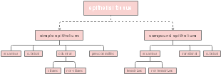
single layer epithelium
single layer squamous epithelium
Observed from the surface, the cells are irregular polygonal, with a smooth surface, jagged cell edges, adjacent cells chimeric to each other, and an oblate nucleus located in the center of the cell. Observed from the vertical section, the cells are very thin and only the part containing the nucleus is slightly thicker.
Endothelium: The single layer of flat epithelium that lines the inner surface of the heart, blood vessels and lymphatic vessels is usually called endothelium. It has a smooth surface and is conducive to blood and lymph flow and material transport.
Mesothelium: The single layer of flat epithelium distributed on the surface of the pleura, peritoneum and pericardium is called mesothelium, which can reduce friction between organs and facilitate organ movement.
single layer cuboidal epithelium
When viewed from the surface, cells appear hexagonal or polygonal; Observed from the vertical section, the cells are cuboidal in shape with a round nucleus located in the center.
It is mainly distributed in renal tubules, thyroid follicles, etc., and has functions such as absorption and secretion.
simple columnar epithelium
When viewed from the surface, cells appear hexagonal or polygonal; In the vertical section, the cells are columnar and the nuclei are oval, mostly located at the base of the cells.
Distributed in the cavities of the stomach, intestines, gallbladder, fallopian tubes, uterus and other organs, it mainly has the functions of absorption and secretion.
pseudostratified ciliated columnar epithelium
It is generally composed of columnar cells (mostly), spindle cells, cone cells, and goblet cells. Observed from the vertical section, because the cells are of different heights and the nuclei are not on the same plane, they appear to be stratified under a light microscope, but the basal surface of all cells is attached to the basement membrane; there are a large number of cilia on the free surface.
This kind of epithelium is mainly distributed in the pharynx, larynx, trachea, bronchi, Eustachian tube, tympanic cavity and other cavity surfaces, and mainly plays a protective role.
stratified epithelium
Stratified squamous epithelium
The surface cells are flat and scaly. When viewed in a vertical section, the layer close to the basement membrane is called the basal layer. It is a stem cell with the ability to proliferate and differentiate. The middle layer is multi-layered polygonal cells, and the surface layer is multi-layered flat cells.
The connection between the epithelium and the deep connective tissue is uneven, which not only ensures the supply of nutrients to the epithelial tissue but also makes the connection stronger.
The epithelial cells distributed on the surface of the skin have no nuclei and contain a large amount of keratin. The cells are dry and hard and can fall off. They are called keratinized stratified squamous epithelium.
The superficial cells distributed in the oral cavity, esophagus and vaginal cavity are living cells with nucleation, containing less keratin, and are called non-keratinized stratified squamous epithelial cells.
Stratified columnar epithelium
It is composed of multiple layers of cells, with a superficial layer of neatly arranged columnar cells and a deep layer of one or several layers of polygonal cells.
This type of epithelium is mainly found in the palpebral conjunctiva, the male urethra and the large ducts of some glands.
metastatic epithelium
Mainly distributed in the renal calyces, renal pelvis, ureters, bladder and other cavities.
Divided into surface layer, middle layer and basal layer. The superficial cap cells are large and thick, in larger round and oval shapes, and some have binucleates.
The characteristic is that the cell layer and morphology can change with the functional status of the organ.
Under a light microscope, the free surface of the cap cells is strongly eosinophilic due to the concentration of cytoplasm, and is called the shell.
Special differentiated structure of epithelial tissue
Specialized structure of the free surface of epithelial cells
1. Microvilli
It is a tiny finger-like protrusion that extends from the cell membrane and cytoplasm to the free surface of the cell.
The striated border of the columnar epithelium of the small intestine and the brush border of the renal tubules seen under light microscopy are both composed of microvilli.
Under an electron microscope, microvilli are about 0.1 μm in diameter, with a cell membrane on their surface, and their axis contains many longitudinal microfilaments.
The outer surface of the cell membrane of microvilli is often covered with a thick layer of cell coat. The cell coat has the functions of adhesion, protection and material exchange. In addition, it is also related to cell surface antigenicity and cell recognition.
Microvilli can expand the surface area of cells and facilitate the cell's absorptive function.
2.Cilia
It is a long protrusion extending from the free surface of epithelial cells and has the function of rhythmic directional swing.
There are two microtubules arranged parallel to its long axis in the center of the cilium, surrounded by 9 groups of doublet microtubules. There is a dense particle at the root called the matrix.
The swing of cilia can push particles, foreign matter, mucus, etc. adhered to the epithelial surface in a direction, so cilia have the function of removing foreign matter and transporting substances.
specialized structures on the sides of epithelial cells
1. Tight connection (occlusive zonules)
Structure: Mostly located at the top; point-like fusion; mesh-like structure
Function: Seals the intercellular space, which not only prevents extracellular macromolecular substances from entering deep tissues through the intercellular spaces, but also prevents the overflow and loss of material components in deep tissues.
2. Middle connection (adhesive tape)
Structure: Located below the tight junction; there is a gap of 15~20nm, and filaments are connected in the middle; there are dense substances attached to the inner surface of the cell membrane and are connected to the terminal mesh filaments
Function: inter-cell adhesion, information transmission, maintaining cell function and shape, and transmitting cell contractility, etc.
3. Desmosomes (focal adhesions)
Structure: There are gaps between the patchy membranes, with sticky substances and filaments inside, and a dense midline; there are attachment plates and tension filaments on the cytoplasmic surface.
Function: Mechanical connection and information transmission between cells (the strongest connection)
4. Gap connection (communication connection)
Structure: Surrounded by 6 connexin molecules, with a lumen of 2 nm in diameter in the center
Function: Exchange small molecule substances and ions; facilitate the conduction of electrical impulses (resistance here is very low)
5. Connection complex (locking embankment)
Two or more of the four types of cell connections exist at the same time
Specialized structure of the basal surface of epithelial cells
1. Basement membrane (basal membrane)
Placode (composed of mucopolysaccharide substances secreted by epithelial cells)
Translucent layer (close to the basal surface of epithelial cells)
Dense layer
Reticulum (matrix and reticular fibers secreted by fibroblasts)
2. Plasma membrane infoldings
They are many cell folds of varying lengths formed by the cell membrane on the basal surface of epithelial cells folding into the cytoplasm and distributed vertically to the basal surface of the cells.
Effectively expands the surface area of the basal surface of cells; facilitates the rapid transport of water, electrolytes and other substances
3. Hemidesmosome
Located at the contact point between the basal surface of epithelial cells and the basement membrane. Its structure is half of a desmosome
Functions to strengthen the connection between epithelial cells and basement membrane
There are many scattered goblet cells between the epithelial cells of the intestinal wall. The nuclei are inverted triangles and deeply stained, near the base of the cells. The top cytoplasm is filled with mucin granules. The mucus formed after secretion can lubricate and protect the epithelium. (The stomach and gallbladder normally do not have goblet cells)