MindMap Gallery Chemical Effects of Electric Current
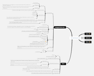
Chemical Effects of Electric Current
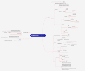
The chemical effects of electric current on chemical substances are widely used in industrial production and scientific research. Current can affect the speed and direction of chemical reactions, and even trigger new chemical reactions. In electrochemical reactions, when the current passes through the electrolyte solution, ion migration and electron exchange occur, which not only change the chemical properties of the substance, but may also trigger electrochemical reactions. These reactions have wide applications in fields such as batteries, electrolysis, and electroplating. In addition, current can also be used for separating and purifying substances, such as electrophoresis and electrocrystallization techniques. Understanding the chemical effects of current helps us better utilize electrochemical processes, optimize industrial processes, and improve production efficiency. At the same time, it also promotes the development of new technologies and provides more possibilities for scientific research. This is a mind map about Chemical Effects of Electric Current. The map contains 6 main branches, which are: What is charge, What is current?, What is conventional current?, Electroplating, Electrolysis, What is Electronic Current?. Each main branch has a detailed description of its sub branches. Suitable for people interested in Chemical Effects of Electric Current.
Edited at 2024-02-02 01:52:26
- Recommended to you
- Outline