MindMap Gallery Classification of Solution Chemistry
Classification of Solution Chemistry
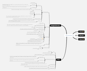
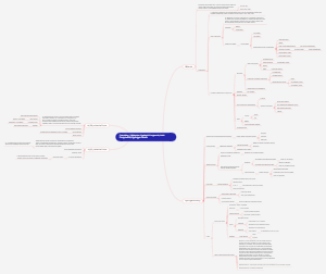
The classification of solution chemistry encompasses solubility, colligative properties, and electrolytes and nonelectrolytes. The solubility section delves into various factors affecting solubility and demonstrates the plotting of solubility curves. The colligative properties section explains phenomena such as osmotic pressure, elevation of boiling point, and depression of freezing point in detail. The electrolytes and nonelectrolytes section clearly distinguishes between electrolytes (strong and weak) and nonelectrolytes, and investigates their ionization and conductivity behavior in solution.
Edited at 2024-12-22 10:13:20- Recommended to you
- Outline