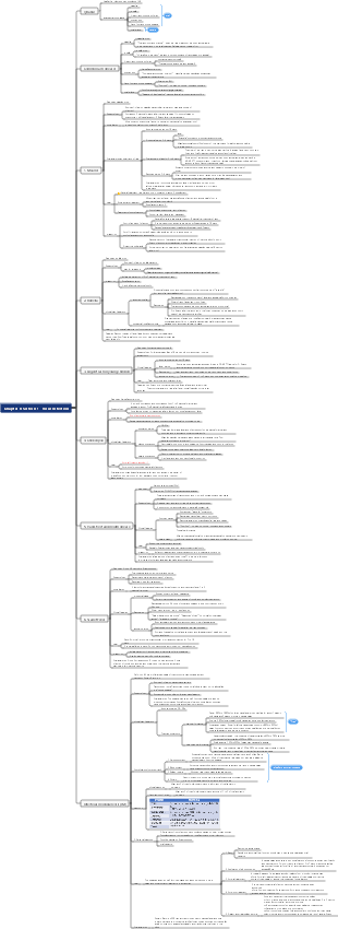
MindMap Gallery Medicine - Neonatal Diseases
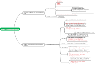
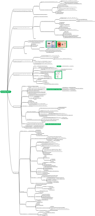
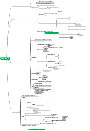
It mainly includes the characteristics and care of normal full-term infants and premature infants, fetal intrauterine growth abnormalities, neonatal hypoxic-ischemic encephalopathy, etc. I hope it can be helpful to everyone.
Edited at 2023-12-09 13:52:01Mappa mentale per l’analisi del controllo della gestione del portata di progetto. Tre sezioni principali: 1. WBS Scope Breakdown – scomposizione gerarchica del lavoro con dettaglio dello Scopo del progetto. 2. Scope Boundary / Exclusions – definizione chiara dei confini del progetto e di ciò che è escluso per evitare scope creep. Strumento ideale per project manager e team di controllo per mantenere allineamento e ridurre rischi.

Questo template, ideato con EdrawMind, è un modello completo per il miglioramento continuo del sistema di gestione della qualità, con una descrizione di circa 500 parole. È strutturato come un diagramma a lisca di pesce, strumento efficace per analizzare le cause principali dei problemi di qualità e definire le leve di controllo necessarie per risolverli. Il processo è organizzato in quattro fasi chiave: la prima è lo standard e la pianificazione, dove si definiscono gli obiettivi di qualità, i criteri di accettazione, i requisiti di prova e le porte di qualità (Quality Gate). La seconda fase analizza i processi e le porte di controllo, per garantire che ogni passaggio del lavoro segua i standard stabiliti. La terza fase riguarda l’esecuzione QA/QC, con la definizione di metodi di prova e ispezione, nonché regole per la gestione dei difetti e delle non conformità. L’ultima fase è il miglioramento e le azioni correttive e preventive (CAPA), insieme a una catena di audit per monitorare l’efficacia delle misure adottate. In basso, una tabella "Quality Gate" permette di tracciare ogni punto di controllo, con criteri specifici, metodi di prova, proprietario e stato di avanzamento. Grazie alla visualizzazione chiara e intuitiva di EdrawMind, questo strumento aiuta il team a identificare le cause root dei problemi di qualità, implementare azioni efficaci e mantenere un ciclo di miglioramento continuo, garantendo la qualità finale del prodotto o servizio.
Questo template, creato con EdrawMind, è un modello di piano di comunicazione ottimizzato, con una descrizione di circa 500 parole. È uno strumento chiave per evitare incomprensioni tra stakeholder, garantire la trasparenza e mantenere il team allineato agli obiettivi del progetto. Il modello è composto da tre elementi fondamentali e interconnessi: la matrice di comunicazione, la cadenza timeline e le regole di comunicazione/SLA. La matrice di comunicazione è una tabella dettagliata dove per ogni pubblico o stakeholder, si definisce il tipo di informazioni da condividere, lo scopo della comunicazione, il canale da utilizzare (email, riunioni, piattaforme di progetto), la frequenza, il proprietario responsabile, il formato e il percorso di escalazione in caso di problemi. La cadenza timeline è una linea temporale che definisce le scadenze delle comunicazioni chiave, garantendo che le informazioni siano condivise in momento opportuno e non si verifichino ritardi o omissioni. Le regole di comunicazione e gli accordi sul livello di servizio (SLA) definiscono le norme formali della comunicazione, come i tempi di risposta, il tono da adottare e le responsabilità di ciascun membro del team. Grazie alla struttura visuale di EdrawMind, questo template permette di pianificare la comunicazione in modo strategico, trasparente e efficiente, riducendo i rischi di cattiva informazione, migliorando la collaborazione e garantendo che tutti gli stakeholder siano informati e coinvolti nel progetto.
Mappa mentale per l’analisi del controllo della gestione del portata di progetto. Tre sezioni principali: 1. WBS Scope Breakdown – scomposizione gerarchica del lavoro con dettaglio dello Scopo del progetto. 2. Scope Boundary / Exclusions – definizione chiara dei confini del progetto e di ciò che è escluso per evitare scope creep. Strumento ideale per project manager e team di controllo per mantenere allineamento e ridurre rischi.
Questo template, ideato con EdrawMind, è un modello completo per il miglioramento continuo del sistema di gestione della qualità, con una descrizione di circa 500 parole. È strutturato come un diagramma a lisca di pesce, strumento efficace per analizzare le cause principali dei problemi di qualità e definire le leve di controllo necessarie per risolverli. Il processo è organizzato in quattro fasi chiave: la prima è lo standard e la pianificazione, dove si definiscono gli obiettivi di qualità, i criteri di accettazione, i requisiti di prova e le porte di qualità (Quality Gate). La seconda fase analizza i processi e le porte di controllo, per garantire che ogni passaggio del lavoro segua i standard stabiliti. La terza fase riguarda l’esecuzione QA/QC, con la definizione di metodi di prova e ispezione, nonché regole per la gestione dei difetti e delle non conformità. L’ultima fase è il miglioramento e le azioni correttive e preventive (CAPA), insieme a una catena di audit per monitorare l’efficacia delle misure adottate. In basso, una tabella "Quality Gate" permette di tracciare ogni punto di controllo, con criteri specifici, metodi di prova, proprietario e stato di avanzamento. Grazie alla visualizzazione chiara e intuitiva di EdrawMind, questo strumento aiuta il team a identificare le cause root dei problemi di qualità, implementare azioni efficaci e mantenere un ciclo di miglioramento continuo, garantendo la qualità finale del prodotto o servizio.
Questo template, creato con EdrawMind, è un modello di piano di comunicazione ottimizzato, con una descrizione di circa 500 parole. È uno strumento chiave per evitare incomprensioni tra stakeholder, garantire la trasparenza e mantenere il team allineato agli obiettivi del progetto. Il modello è composto da tre elementi fondamentali e interconnessi: la matrice di comunicazione, la cadenza timeline e le regole di comunicazione/SLA. La matrice di comunicazione è una tabella dettagliata dove per ogni pubblico o stakeholder, si definisce il tipo di informazioni da condividere, lo scopo della comunicazione, il canale da utilizzare (email, riunioni, piattaforme di progetto), la frequenza, il proprietario responsabile, il formato e il percorso di escalazione in caso di problemi. La cadenza timeline è una linea temporale che definisce le scadenze delle comunicazioni chiave, garantendo che le informazioni siano condivise in momento opportuno e non si verifichino ritardi o omissioni. Le regole di comunicazione e gli accordi sul livello di servizio (SLA) definiscono le norme formali della comunicazione, come i tempi di risposta, il tono da adottare e le responsabilità di ciascun membro del team. Grazie alla struttura visuale di EdrawMind, questo template permette di pianificare la comunicazione in modo strategico, trasparente e efficiente, riducendo i rischi di cattiva informazione, migliorando la collaborazione e garantendo che tutti gli stakeholder siano informati e coinvolti nel progetto.
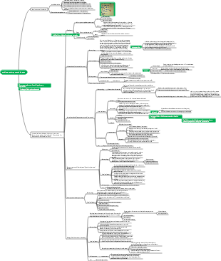
neonatal diseases
Overview
Gestational age classification
Expired≥42
Full term 37≤x<42
Premature birth <37
Weight classification
Normal weight 2500≤x≤4000g
Low body weight <2500g
Extremely low body weight <1500g
Ultra low weight <1000g
Relationship between birth weight and gestational age
Suitable for gestational age infants → bw (birth weight) is 10%-90% of the average weight of the same gestational age
Small for gestational age → bw below 10% of average weight
Large for gestational age → bw is above 90% of average weight
Supplement: perinatal period (gestational age 28 weeks - 7 days after birth)
Characteristics and care of normal term infants and premature infants
Exterior
Full term
Less vellus hair, breast nodules >4mm, nails extending beyond fingertips, and many foot prints
premature birth
Excessive vellus hair, breast nodules <4 mm, nails less than fingertips, and few foot prints
Physiological characteristics
respiratory system
Full term → Fetal lungs are filled with fluid. After delivery, the alveolar cells switch from secretion to absorption → the fluid decreases → squeezed during delivery → the fluid can be discharged through the mouth and nose
premature birth
Hypoplasia of respiratory center → insensitive to hypoxia and hypercapnia
Fewer alveoli → less gas exchange
The ventilator is underdeveloped → shallow and fast breathing
Irregular breathing or even apnea
circulatory system
Premature birth → there may be an open ductus arteriosus
The normal heart rate is 120-140
Breathing is 35-45
digestive urinary system
Full term
Digestive tract → Because the esophageal sphincter is relaxed and the stomach level → prone to galactorrhea and vomiting of milk
Defecation → Start defecation within 24 hours after birth (dark green) and finish in 2-3 days
Urination → Urination within 24 hours after birth, up to 20 times a day within a week
Immature kidney function → edema may occur
nervous system
Spinal cord → lumbar puncture at 4-5 space (the end of the spinal cord is at the lower edge of 3-4 space)
reflection
Primitive reflex (normally disappears after a few months)
feeding reflex
sucking reflex
grasping reflex
hug reflex
Pathological reflexes (appear as normal conditions)
kerning's sign
Babinski sign
chvostek sign
ps: The abdominal wall and cremaster reflexes are unstable, and occasionally there may be paroxysmal ankle clonus.
Body temperature (newborns are prone to hypothermia)
Imperfect body temperature regulation
Poor skin keratinization
Thin subcutaneous fat
Large body surface area
brown fat deficiency
immune system
Placenta related
igG can pass through the placenta (higher content in the body)
igA and igE cannot cross the placenta → susceptible to bacterial (gram-positive bacterial infection)
Immune cells → Low T cell function is the main cause of incompetent immune response
special physiological state
physiological jaundice
horse teeth, mantis mouth
Breast enlargement, false menstruation
neonatal erythema
Milia
Term and premature infant care
Respiratory management (be careful to avoid routine oxygen inhalation → high concentration of oxygen can cause retinopathy) → also the most serious complication of oxygen therapy for premature infants
intrauterine growth abnormalities
Intrauterine growth retardation and small for gestational age
Cause
Maternal factors (malnutrition, multiple parities, placental factors, eclampsia, parental short stature)
Fetal factors (intrauterine infection, Down syndrome, meowing syndrome, multiple births)
complication
Perinatal asphyxia (long-term lack of oxygen in the womb)
congenital malformation
Hypoglycemia
meconium aspiration syndrome
Polycythemia—high viscosity
Large for gestational age
Cause
Physiological factors (parents are tall and eat a lot during pregnancy)
Pathological factors (pregnant mother has diabetes, RH blood group incompatibility, congenital heart disease)
clinical manifestations
asphyxia
intracranial hemorrhage
Various birth injuries (nerve damage, liver rupture)
Neonatal asphyxia (mostly continued intrauterine distress)
Cause
Maternal factors (cardiopulmonary insufficiency, high blood pressure, drug abuse and smoking)
Placental factors (placenta previa, placental abruption)
Umbilical cord factors (umbilical cord prolapse, neck wrapping, knotting)
fetal factors
premature infant macrosomia
Congenital malformations (pulmonary hypoplasia, congenital heart disease)
intrauterine infection
airway obstruction
Childbirth factors (cephalopelvic misalignment, uterine atony)
Pathophysiology
When suffocating → normal breathing cannot be established → lung fluid cannot flow out
Hypoxia → decrease in pulmonary surfactant and increase in pulmonary vascular resistance → pulmonary hypertension
Breathing changes
primary apnea
Manifestations (in the early stage of hypoxia, compensatory acceleration of breathing → if no effective treatment → respiratory arrest)
Treatment (removing the cause, clearing the respiratory tract, physical stimulation)
secondary apnea
Performance (hypoxia persists→after several deep wheezing breaths→respiratory arrest)
Treatment (positive pressure ventilation)
clinical manifestations
Fetal distress (fetal heart rate ≥160)
apgarscore
Content (skin color, muscle tone, response to stimulation: bouncing the soles of the feet, inserting a nasal cannula, heart rate, breathing) showing off the muscles and getting emotional after being beaten
score
Normal 10-8
Moderate 4-7
Severe 0-3
treat
Resuscitation Program (ABCDE)
aClear the respiratory tract
bEstablish breathing
cMaintain normal circulation
dDrug therapy
e-assessment
Resuscitation steps
(Quick assessment) Is it full term? Is the amniotic fluid clear? Is there any crying? Is the muscle tone good?
No (preliminary assessment)
Keep warm → Position yourself → Clear the airway → Dry the body → Stimulate and induce spontaneous breathing → Assess whether the heart rate is <100 beats, apnea, wheezing breathing
Yes (positive pressure ventilation: because of secondary apnea)
Effective (heart rate increases rapidly ≥100 with spontaneous breathing)
Invalid (no spontaneous breathing, heart rate <100)
If the heart rate is still <60 after 30 seconds → add external chest compressions (depth is 3/1 of the front and rear chest diameter) and endotracheal intubation at the same time
Effective (routine care)
Ineffective (adrenaline medication)
If the chest rises poorly → just intubate the trachea
None (routine care)
Yes (for routine care)
neonatal hypoxic-ischemic encephalopathy
Cause
Hypoxia (Core)
Lung disease, hypoxia, anemia
Pathogenesis
cerebral blood flow
The basal ganglia, brainstem, and thalamus are the first to ensure blood supply
Damage to parasagittal areas and white matter of cerebral cortex
The vulnerable area in term infants is in the parasagittal area of the brain (cerebral cortex), and in premature infants is in the ventricular white matter area. (Pig food jujube white)
Changes in brain tissue metabolism (oxygen consumption and metabolism in newborns are higher than in adults)
pathology
Cerebral edema (early major change)
clinical manifestations
Consciousness, muscle tone, primitive reflexes, convulsions, disease course, prognosis
Mild (consciousness → agitation, hugging reflex → active, sucking reflex → normal, symptoms disappear after 72h (3 days) without sequelae, pupils are dilated)
Moderate (consciousness → drowsiness, symptoms disappear within 14 days, some with sequelae, miosis)
Severe (consciousness → coma, all reflexes disappear, death may occur within days to weeks, pupils are fixed and light reflex disappears)
Auxiliary inspection
brain imaging test
B-ultrasound (preferred) ps: insensitive to the parasagittal area
CT (suitable time is 4-7 days after birth)
MRI magnetic resonance (sensitive to parasagittal area)
Electroencephalogram (can determine the extent of damage)
diagnosis
Have a history of intrauterine distress
Fetal heart rate <100 for more than 5 minutes
Amniotic fluid third degree pollution
History of asphyxia during delivery/severe asphyxia at birth (apgar score ≤ 3)
Changes in consciousness/muscle tone/abnormal reflexes after birth
treat
supportive care
Get up early → control symptoms and maintain internal environment
Mid-term→guaranteed perfusion
Late stage → functional recovery
drug
Control convulsions
Normal function→phenobarbital
Hepatic insufficiency→phenytoin
Treat cerebral edema
Furosemide is preferred
Mannitol and corticosteroids are contraindicated
neonatal intracranial hemorrhage
Etiology and pathogenesis
Premature birth (when the brain is still developing)
Ischemia and hypoxia (hypercapnia during asphyxia)
Physical injury (fetal malposition, macrosomia, premature delivery)
Clinical manifestation classification
Periventricular - intraventricular hemorrhage (more common in premature infants) → rapid increase in head circumference, full anterior fontanelle, cranial suture separation, intellectual motor impairment
Primary subarachnoid hemorrhage (more common in newborns) → good prognosis
Cerebral parenchymal hemorrhage (more common in term infants) (most common type)
diagnosis
CT and MRI? (MRI is the first choice)
Neonatal meconium aspiration syndrome (respiratory mechanical obstruction, chemical inflammation of lung tissue, respiratory distress after birth)
Cause/Pathology/Physiology
Meconium aspiration (high incidence of macrosomia)
Uneven airway obstruction (meconium mechanically blocks the airway) (atelectasis, emphysema, and normal alveoli coexist)
Atelectasis→Cause: The small airways of some alveoli are completely blocked (completely blocked) by larger meconium particles
Emphysema → Cause: Some alveolar small airways are partially blocked by some sticky meconium particles → forming a valve (partial blockage)
Normal alveoli → normal alveoli become more compensatory due to obstruction of other alveoli
Chemical inflammation of lung tissue→Meconium stimulation→Chemical inflammation of lung tissue Interstitial emphysema
pulmonary hypertension
diagnosis
Common term infants and macrosomia
Multiple history of intrauterine distress and birth asphyxia
Symptoms are related to the amount of amniotic fluid (meconium contained in it) inhaled
clinical manifestations
Aspiration of amniotic fluid mixed with meconium → gold standard (meconium can be seen by aspiration of the glottis and trachea)
Imaging manifestations (extensive emphysema in both lungs, nodular high-density shadows)
Neonatal respiratory distress syndrome, also known as hyaline membrane disease (deficiency of pulmonary surfactant ps) (more common in premature infants, the younger the gestational age, the higher the incidence)
ps component → type II alveolar epithelial cells (lecithin/sphingomyelin ratio → used as an indicator to evaluate fetal lung maturity)
Cause
Premature birth (the younger the gestational age, the less PS is secreted and synthesized)
baby of diabetic mother
Cesarean section (lack of uterine contractions, weak hormone response → affecting synthesis)
Mechanism (decreased alveolar surfactant → increased alveolar surface tension) → decreased lung compliance, increased airway resistance, decreased ventilation/blood flow (increased tension, decreased compliance, decreased residual volume ventilation)
clinical manifestations
Premature babies are more common
Symptoms include (shortness of breath, moaning, bruising, three concave signs)
complication
Patent ductus arteriosus (the original disease has improved but suddenly increased oxygen demand occurs, metabolic acidosis)
Auxiliary inspection
Blood gas analysis (arterial oxygen partial pressure decreases and carbon dioxide partial pressure increases)
x-ray
ground glass
dendritic shadows (air bronchial sign)
white lung
treat
Mechanical ventilation and application of pulmonary surfactant-based closure of the ductus arteriosus
How to use the active substance → Shake the drug well and inject it into the lungs through the trachea
Mechanical ventilation method mode: ippv peep
If it is early →continuous positive airway pressure
Prevention (if the pregnancy is less than 34 weeks, prenatal hormones will be given before delivery)
Supplement (wet lung) → more common in full-term infants and cesarean section infants
Brief description: Neonates have temporary dyspnea and transient shortness of breath after birth, but the symptoms disappear quickly and the prognosis is good.
Cause
increased fluid in the lungs
Increased fluid in the lungs and insufficient lymphatic drainage from the lungs
The alveoli retain more fluid, which affects gas exchange and causes difficulty breathing.
clinical manifestations
Most cases of respiratory acceleration (>60 times/min) appear within 6 hours after birth.
Weak cry, cyanosis, slight moaning, nasal fluttering, three concave signs, rapid breathing
Raised PaCO2 and acidosis are uncommon
diagnosis
Breathing is mostly normal at birth, but tachypnea and cyanosis develop within 6 hours after birth.
Pulmonary signs are not obvious, only decreased breath sounds or rough crackles
Shortness of breath usually disappears within 24 hours
Image (X check spotty cloud shadow)
neonatal jaundice
Physiological (most bilirubin comes from senescent red blood cells)
physiological jaundice
Full-term infants <221 (12.9)
Premature infants <256 (15)
Unconjugated bilirubin is the most common
Unconjugated bilirubin combined with albumin → non-toxic
Free unconjugated bilirubin can pass the blood-brain barrier
Metabolic characteristics
Excessive bilirubin production
Newborns produce more bilirubin than adults
Newborn red blood cells have short lifespan
Hemoglobin is broken down twice as fast as adults
Insufficient ability of plasma albumin to bind bilirubin
Poor processing capacity of liver cells
Characteristics of enterohepatic circulation
High enzyme activity → high enterohepatic circulation → more intestinal recycling
Slow excretion of meconium → excessive bilirubin absorption
Low albumin and high unconjugated bilirubin
Classification of jaundice
Physiological jaundice (appears 2-3 days after birth and disappears 2 weeks after birth)
Pathological jaundice (appears within 1 day of birth)
Preterm infants lasting >4 weeks
Term infants last >2 weeks
Cause
Too much bilirubin production
Increased red blood cells
Hemolysis
Infect
Increased enterohepatic circulation
breast milk
Breastfeeding-related jaundice (within one week → insufficient feeding caloric and fluid intake → delayed defecation → elevated serum bilirubin)
Breast milk jaundice (breast milk causes increased enterohepatic circulation → jaundice) (can get better after stopping breastfeeding for 1-2 days)
Hepatic bilirubin metabolism disorder
Bile excretion disorder
Hypoxia will worsen
Hemolytic disease of the newborn (ABO incompatibility is more common, RH incompatibility is less common)
Cause
ABO hemolysis (most common, multiple first birth, mother must be type O)
The mother is O and the fetus is A or B (the type O mother has been stimulated by A or B blood group substances → plants/parasites/vaccines before her first pregnancy) to produce A or B antibodies.
The mother is AB and the fetus is O
RH hemolysis (multiple second pregnancy, mother is rh negative, child is rh positive) (rh positive blood has been transfused in the past) → rh antibodies can only be stimulated by human red blood cells, so the first pregnancy will not occur
Pathophysiology (excessive serum unconjugated bilirubin due to hemolysis → bilirubin encephalopathy)
clinical manifestations
jaundice
Most rh hemolysis → appears 24 hours after birth
abo hemolysis → appears in 2-3 days
anemia
Hepatosplenomegaly
rh more
abo generally no
complication
Bilirubin encephalopathy (kernicterus)
Warning period - the first stage (drowsiness, low reaction, weakened reflexes, screaming and vomiting)
Spasmodic phase—second phase (convulsions, increased muscle tone, opisthotonus)
Recovery period—the third period (response improves, convulsions decrease)
Sequelae Phase—Phase 4
Athetosis (involuntary, purposeless movements)
eye movement disorder
Hearing impairment
Enamel dysplasia
cerebral palsy
laboratory tests
Blood type test (preferred)
Check for hemolysis
Sensitized blood cells and blood group antibody determination
Modified direct antiglobulin test (also called coombs test) → measure rh hemolysis
Antibody release test → measure abo hemolysis
treat
prenatal treatment
Neonatal period therapy
Phototherapy (phototherapy makes unconjugated bilirubin isomerize → easily soluble in water → can not pass through the liver → be directly excreted through bile and urine → reduce serum unconjugated bilirubin)
The cause is unknown, but symptoms are mild and can be used
medical treatement
Supply albumin
Correct acid
exchange transfusion therapy
Indications
Total bilirubin>342
severe hemolysis
Bilirubin encephalopathy
method
If it is mother o, child a or b has abo hemolysis → use mixed blood of ab plasma and o red blood cells
Volume of blood transfusion: generally twice that of the patient
Neonatal infectious diseases (common pathogens: bacteria and viruses)
neonatal sepsis
Etiology and pathogenesis
Pathogenic bacteria (staphylococci are the most common) (followed by Escherichia coli)
Characteristics of neonatal immune system
non-specific immune system
specific immune system
The IgG in newborns mainly comes from the mother → the smaller the gestational age, the lower the IgG content → so premature babies are more susceptible to infection
The molecular weight of igA and igM is large → cannot pass through the placenta → the content of the fetus is low → susceptible to infection by Gram-negative bacilli
clinical manifestations
Early/late hair
early onset
Onset within 7 days of birth, usually before/before birth
Mostly caused by maternal vertical transmission (placenta, birth canal, breastfeeding)
Mainly Escherichia coli (G-)
late hair style
Onset of illness after 7 days
Mostly caused by horizontal transmission (skin, digestive tract, respiratory tract)
Mainly Staphylococcus aureus
Early symptoms
Jaundice (rapidly worsening jaundice)
Hepatosplenomegaly
Bleeding tendency (skin petechiae)
Shock (skin marbling)
Auxiliary inspection
Bacteriological examination
Blood culture (preferred) → C-reactive protein
cerebrospinal fluid/urine culture
subtopic
treat
Early onset → third generation cephalosporins
Late onset→oxacillin
If resistant →vancomycin
Neonatal infectious pneumonia (common ones include: Escherichia coli, Klebsiella pneumoniae)
Cause
Intrauterine infectious pneumonia (mainly virus)
Infectious pneumonia during delivery (premature rupture of membranes, prolonged labor, loose disinfection)
infection after birth
clinical manifestations
Most children have a history of asphyxiation after birth, with rough and moist rales on lung auscultation, shortness of breath, cyanosis, and three concave signs.
Viral pneumonia x-image → interstitial pneumonia changes
Bacterial pneumonia x-image → manifestations of bronchopneumonia
Image (patchy exudation)
neonatal tetanus
What can cause milk overflow
Gastroesophageal reflux (transverse stomach)
Pylorospasm (developed pyloric sphincter)
Esophageal atresia
Improper feeding
Neonatal digestive tract supplement
The stomach is in a transverse position
Underdeveloped cardiac sphincter → prone to galactorrhea
Well developed pyloric sphincter
Newborns classified according to age
Early neonates (within one week after birth)
Late neonate (2 weeks to 4 weeks after birth)
Metabolism and excretion disorder supplements
GILbert syndrome (hepatic bilirubin uptake disorder) (hepatic glucuronosyltransferase utilization disorder) Missed shot
Crigler-Najjar syndrome (congenital glucuronosyltransferase deficiency) Ronaldo is missing the ball
Dubin-Johnson syndrome, hepatocellular secretion and excretion disorder, feces is blocked
Kernicterus = Bilirubin Encephalopathy
Relaxation of muscle tone is scored as 0 points, slight flexion of the limbs is scored as 1 point, and limb activity is scored as 2 points.
ps: If the heart rate is not 0 points, <100 is 1 point, and >100 is 2 points
Newborns (umbilical cord ligation to within 28 days after birth)
HiE also known as ischemic hypoxic encephalopathy
NOTE: Neonatal respiratory distress syndrome = hyaline membrane disease