MindMap Gallery respiratory system
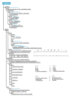
This is a mind map about the respiratory system. The respiratory system is the general name for a series of organs that exchange gases between the human body and the outside air. It is rich in content, summarized in key points, clear in structure, and complete in system! Very worth learning!
Edited at 2024-11-01 11:41:41This is a mind map about the annual work plan of the three pillars of human resources. The main contents include: strategic human resources planning, talent recruitment and allocation, employee performance management, employee training and development, employee relationships and communication, employee welfare and care, human resources information system construction, regulatory compliance and risk management, and organizational culture construction.
This is a mind map for the diagnosis and treatment of acute cerebral hemorrhage in patients with hemodialysis. The annual incidence of acute cerebral hemorrhage in patients with hemodialysis is (3.0~10.3)/1000, and the main cause is hypertension. Compared with non-dialysis patients, the most common bleeding site is the basal ganglia area, accounting for 50% to 80%; but the bleeding volume is large and the prognosis is poor, and the mortality rate is 27% to 83%. Especially for patients with hematoma >50ml, hematoma enlarged or ventricular hemorrhage on the second day after onset, the prognosis is very poor.
The logic is clear and the content is rich, covering many aspects of the information technology field. Provides a clear framework and guidance for learning and improving information technology capabilities.
This is a mind map about the annual work plan of the three pillars of human resources. The main contents include: strategic human resources planning, talent recruitment and allocation, employee performance management, employee training and development, employee relationships and communication, employee welfare and care, human resources information system construction, regulatory compliance and risk management, and organizational culture construction.
This is a mind map for the diagnosis and treatment of acute cerebral hemorrhage in patients with hemodialysis. The annual incidence of acute cerebral hemorrhage in patients with hemodialysis is (3.0~10.3)/1000, and the main cause is hypertension. Compared with non-dialysis patients, the most common bleeding site is the basal ganglia area, accounting for 50% to 80%; but the bleeding volume is large and the prognosis is poor, and the mortality rate is 27% to 83%. Especially for patients with hematoma >50ml, hematoma enlarged or ventricular hemorrhage on the second day after onset, the prognosis is very poor.
The logic is clear and the content is rich, covering many aspects of the information technology field. Provides a clear framework and guidance for learning and improving information technology capabilities.
respiratory system
Division of respiratory system
Upper respiratory tract: nose, pharynx; larynx
Lower respiratory tract: trachea, main bronchi, and branches at all levels within the lungs
The pharynx belongs to both the digestive system and the respiratory system
nose
external nose
Root of nose, dorsum of nose, tip of nose, wings of nose, nostrils
The nasal bones and cartilage serve as a scaffold, covered by skin on the outside and mucous membrane on the inside.
Cartilaginous skin is rich in sebaceous and sweat glands and is a favorite site for acne, rosacea and boils.
nasal cavity
nasal vestibule
Located in the front and lower part of the nasal cavity, front and lower part of the nasal region
Made of cartilage covering the skin
Rich in sebaceous and sweat glands. Have nose hair
Boils are prone to occur in areas
proper nasal cavity
Attachment: nasal mucosa
olfactory area
Pale in color and contains a large number of olfactory cells
The inner surface of the superior turbinate, the part of the nasal septum corresponding to the superior turbinate, and the mucosa on the roof of the nasal cavity above them
breathing zone
The remaining part of the mucosa except the olfactory region is continuous with the mucosa of the inner surface of the paranasal sinuses.
Pink in color, rich in blood vessels and nasal glands, it can warm, moisturize and purify the air
Bone, cartilage, mucous membrane, skin composition
Go forward through the nostrils to connect to the outside world, and go backward through the posterior nostrils to connect to the pharynx.
The nasal septum separates the nasal cavity and forms the inner wall of the nasal cavity that is prone to bleeding.
lateral wall
The turbinates arranged from top to bottom: superior turbinate, middle turbinate, inferior turbinate
Nasal meatus arranged from top to bottom, superior meatus, middle meatus, inferior meatus
Structure of the middle meatus, hiatus semilunaris, ethmoid infundibulum, ethmoid bleb
The middle meatus contains most of the openings of the paranasal sinuses
There is an opening of the nasolacrimal duct in the front part of the inferior meatus
The fossa between the posterior and superior aspects of the superior turbinate and the body of the sphenoid is called the sphenoethmoidal recess
paranasal sinuses
maxillary sinus
Maxillary body, average volume 13ml
Anterior wall, posterior wall, upper wall, lower wall, medial wall
It opens in the middle meatus (behind the semilunar hiatus). Because the sinus opening is higher than the sinus floor, it is most susceptible to infection and effusion.
frontal sinus
Located deep to the eyebrow arch. One on each side
Triangular pyramid, base down, tip up
Open into middle meatus
sphenoid sinus
Located in the upper and posterior part of the nasal cavity, within the body of the sphenoid bone
The septum is divided into left and right chambers
Opening into sphenoethmoidal recess
ethmoid sinus
Within the ethmoid labyrinth
Spongy small air cells, ranging from 3 to 18, divided into three groups: front, middle and back
The anterior and middle ethmoid sinuses open into the middle meatus, and the posterior ethmoid sinuses open into the superior meatus.
Air cavities in the skull around the nasal cavity, there are 4 pairs
Opens into the nasal cavity, covers the inner surface with mucosa, and is continuously connected with the nasal mucosa
Warms, moistens and purifies the air, and resonates with pronunciation
pharynx
throat
Alignment C3~C6
laryngeal cartilage
Thyroid cartilage
Made up of two quadrilateral cartilage plates
The front edges of the two plates are connected to form the front horn, and the upper end of the front horn protrudes forward and is called the Adam's apple.
cricoid cartilage
Located below the thyroid cartilage
cricothyroid joint
cricoarytenoid joint
arytenoid cartilage
Located on both sides of the upper edge of the circumferential cartilage plate
One on each side
Epiglottis cartilage: located on the inner surface of the anterior corner of the thyroid cartilage and behind the body of the hyoid bone
connect
key joint
The articular surface of the cricoid cartilage and the inferior angle of the thyroid cartilage are composed of
The thyroid cartilage moves about the coronal axis passing through the cricothyroid joint. Tight or loose vocal cords
cricoarytenoid joint
Cricoid articular surface and spoon-shaped cartilage base
The arytenoid cartilage can rotate internally or externally around the vertical axis, narrowing or opening the loudspeaker.
elastic cone
Conical elastic membrane, also known as surround acoustic membrane
It originates from the inner surface of the anterior horn of the thyroid cartilage, and is fan-shaped and attached posteriorly and downwardly between the upper edge of the cricoid cartilage and the vocal cord process of the arytenoid cartilage.
The upper edge is free and thickened (called the vocal ligament), and the anterior and lower part is thickened (called the median cricothyroid ligament)
square membrane
It originates from the inner surface of the anterior horn of the thyroid cartilage and both sides of the epiglottic cartilage, and connects posteriorly to the anteromedial surface of the arytenoid cartilage.
The upper edge is free and thickened (covered by the arytenoid perineal wall), and the lower part is thickened (called the vestibular ligament)
thyrohyoid membrane
Connected between the upper edge of the thyroid cartilage and the hyoid bone
laryngeal cavity
From the larynx to the lower edge of the cricoid cartilage
Throat
It is composed of the upper edge of the epiglottis, the spoon-shaped epiglottic fold, and the interarytenoid notch.
Two pairs of mucosal folds
Above: vestibular folds, the middle fissure is the vestibular fissure
Below: the vocal fold, covering the vocal cord muscles and vocal ligaments, together they are called the vocal folds. cleft glottis
The glottis cleft is the narrowest part of the larynx. The glottis cleft and the vocal cords are collectively called the glottis.
cleft glottis
The cleft between the vocal folds on both sides and the base of the arytenoid cartilage to the vocal cord process
The anterior 2/3 is located between the vocal folds, called the intermembranous part, and the posterior 1/3 is located between the arytenoid cartilages, called the intercartilage part.
laryngeal cavity division
Laryngeal vestibule: above the level of the vestibular folds
Middle laryngeal cavity: between the plane of the vestibular fold and the plane of the vocal fold, also called the laryngeal chamber, which is the smallest part of the three parts of the laryngeal cavity.
Subglottic space: from below the level of the vocal fold to the lower edge of the cricoid cartilage
laryngeal muscles
cricothyroid muscle and thyroarytenoid muscle
Contraction of the cricothyroid muscle tenses the vocal cords
The contraction of the thyroarytenoid muscle relaxes the vocal cords
Posterior cricoarytenoid muscle and lateral cricoarytenoid muscle
The retroarytenoid muscles contract and the glottis opens wide
The cricoarytenoid muscles contract and the glottis narrows
It is both the passage of the respiratory tract and the organ of speech.
Use the throat opening to open the larynx and pharynx, and lower the inhalation tube
Laryngeal cartilage, connections, laryngeal muscles, mucous membrane composition
trachea, main bronchi
It is composed of 14 to 17 C-shaped cartilage rings and interannular ligaments.
The trachea is divided into left and right main bronchi at the sternal angle plane, and the bifurcation points are called tracheal bifurcations.
There is a tracheal ridge on the inner surface of the tracheal bifurcation, slightly to the left, which is an important sign of bronchoscopy.
Foreign objects entering from the trachea may easily fall into the right main bronchus
From the lower edge of the cricoid cartilage to the tracheal bifurcation
Divided into neck and chest
Composed of tracheal cartilage, connective tissue, smooth muscle, and mucosa
The thyroid isthmus is located in front of the 2nd to 4th tracheal cartilages.
The tracheotomy site is between the 3rd and 4th tracheal cartilage
The left main bronchus is thin, long, and horizontal; the right main bronchus is thick, short, and steep.
lung
Located in the chest, on both sides of the mediastinum
The right lung is wider and shorter than the left lung because of the liver below the diaphragm.
There is a notch in the lower part of the front edge of the left lung
hilum
Located in the center of the mediastinum of the lungs
It is the portal for entry and exit of main bronchi, pulmonary arteries, veins, lymphatic vessels, nerves, etc.
The above structures are wrapped by connective tissue and connect the lungs to the mediastinum, which are called pulmonary roots.
arrangement
From front to back: superior pulmonary vein, pulmonary artery, bronchi
top down
Left lung: pulmonary artery, left main bronchus, inferior pulmonary vein
Right lung: upper lobe bronchi, pulmonary artery, inferior pulmonary vein
Attachment: The front and bottom are the pulmonary veins
Trachea - main bronchi - lobar bronchi - segmental bronchi - bronchopulmonary segments
Light red, spongy, soft and elastic
The apex of the lung is blunt and round, protruding into the base of the neck through the upper thoracic opening, two to three centimeters above the inner 1/3 of the clavicle.
The bottom of the lungs is close to the diaphragm, forming a half-moon depression.
The rib surface is the outer surface, which is in contact with the side of the thorax and the anterior and posterior walls.
The mediastinal surface is the medial surface, connected to the mediastinum, and the oval depression in the middle is the hilum.
Lung root
The entry and exit structures that are surrounded by connective tissue are called pulmonary roots. The arrangement relationship of the main structures in the lung root is divided into static, dynamic and branch from front to back.
Pleura and pleural cavity
The visceral pleura and parietal pleura migrate to each other to form two pleural spaces called pleural cavities Characteristics of the pleural cavity: potential, airtight, negative pressure
parietal pleura
costal pleura
diaphragm pleura
mediastinal pleura
Pleural roof: the highest point is 1 to 4 cm above the medial 1/3 of the clavicle.
visceral pleura
pleural recess
In the pleural cavity, where even the edges of the lungs cannot reach during deep breathing
The costophrenic recess is the lowest-located and largest-capacity part, and inflammatory exudate in patients with pleurisy tends to accumulate here.
The pleura covers the surface of the lungs, turns at the roots of the lungs, and lines both sides of the mediastinum. It is a continuous layer of serous membrane above the diaphragm and inside the chest wall.
pleural cavity
The potential gap formed by the migration of the visceral and parietal pleura at the lung root
There is one on the left and one on the left, which are not connected to each other, are potentially airtight, and present negative pressure, which is conducive to lung expansion.
There is a small amount of slurry inside to reduce friction
Pleural damage causes the negative pressure in the pleural cavity to disappear, resulting in atelectasis, which is called pneumothorax and pleural effusion.
mediastinum
A general term for all the organs, structures and connective tissues between the mediastinal pleura on both sides. It is a sagittal area that is narrow at the top and wide at the bottom, and narrow at the front and back.
The mediastinum is divided into upper mediastinum and lower mediastinum