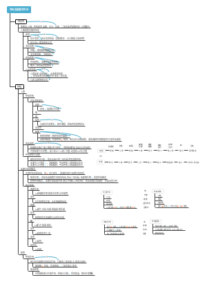
MindMap Gallery respiratory system
This is a mind map about the respiratory system that provides a detailed overview of the structure and function of the respiratory system, from the upper respiratory tract to the lower respiratory tract, to key components such as the lungs and pleura, all presented clearly.
Edited at 2024-11-12 20:09:51これは、「Amazon Reverse Working Method」「Amazon Reverse Working Method」に関するマインドマップです。それは、Amazonの成功の秘密を明らかにし、実用的な作業方法と管理の原則を提供し、Amazon文化を理解し、仕事の効率と創造性を向上させたい読者にとって大きな参照価値です。
Azure BlobストレージにおけるMicrosoftの顕著な進歩とイノベーション、特にChatGptの作成者であるOpenaiの巨大なコンピューティングニーズを効果的にサポートする方法に焦点を当てています。 Azure Blobストレージ製品管理チームのJason Valerieは、JakeとDeverajaと協力して、Azure BlobストレージがOpenaiの大規模なモデルトレーニング、処理データ、ストレージをexebbitレベルまでに行う上で重要な役割を果たしました。議論には、AIワークロードのスケーリングスーパーコンピューターが直面している課題と、地域ネットワークゲートウェイを接続するデータセンターなどのアーキテクチャソリューション、および動的ストレージ容量の拡張を可能にする拡張アカウントの導入が含まれます。技術的な側面は、チェックポイントのメカニズム、大規模なデータ処理、革新的なブロブビューと階層的な名前空間、グローバルデータモビリティ機能をカバーし、Microsoftのグローバルネットワークインフラストラクチャを戦略的に利用して効率的なデータ送信を可能にします。この会話は、高度なAIの研究開発に強力でスケーラブルで効率的なストレージソリューションを提供するというマイクロソフトのコミットメントを完全に示しています。
これは、主にオブジェクト状態の変化、熱エンジン、内部エネルギー、熱比熱容量、温度スケールを含む、熱に関するマインドマップです。紹介は詳細であり、説明は包括的です。
これは、「Amazon Reverse Working Method」「Amazon Reverse Working Method」に関するマインドマップです。それは、Amazonの成功の秘密を明らかにし、実用的な作業方法と管理の原則を提供し、Amazon文化を理解し、仕事の効率と創造性を向上させたい読者にとって大きな参照価値です。
Azure BlobストレージにおけるMicrosoftの顕著な進歩とイノベーション、特にChatGptの作成者であるOpenaiの巨大なコンピューティングニーズを効果的にサポートする方法に焦点を当てています。 Azure Blobストレージ製品管理チームのJason Valerieは、JakeとDeverajaと協力して、Azure BlobストレージがOpenaiの大規模なモデルトレーニング、処理データ、ストレージをexebbitレベルまでに行う上で重要な役割を果たしました。議論には、AIワークロードのスケーリングスーパーコンピューターが直面している課題と、地域ネットワークゲートウェイを接続するデータセンターなどのアーキテクチャソリューション、および動的ストレージ容量の拡張を可能にする拡張アカウントの導入が含まれます。技術的な側面は、チェックポイントのメカニズム、大規模なデータ処理、革新的なブロブビューと階層的な名前空間、グローバルデータモビリティ機能をカバーし、Microsoftのグローバルネットワークインフラストラクチャを戦略的に利用して効率的なデータ送信を可能にします。この会話は、高度なAIの研究開発に強力でスケーラブルで効率的なストレージソリューションを提供するというマイクロソフトのコミットメントを完全に示しています。
これは、主にオブジェクト状態の変化、熱エンジン、内部エネルギー、熱比熱容量、温度スケールを含む、熱に関するマインドマップです。紹介は詳細であり、説明は包括的です。
respiratory system (for gas exchange)
Respiratory tract (breathing tube)
upper respiratory tract
nose and throat
nose
nasal cavity
It is divided into two imperfectly symmetrical left and right cavities by the nasal septum. Nasal septum: vertical plate of ethmoid bone, vomer bone, septal cartilage covered with mucosa The mucosa in the front and lower part of the nasal septum contains a rich capillary network, and epistaxis is prone to occur in this area, which is called Little's area.
nasal vestibule
proper nasal cavity
The bottom wall of the nasal cavity is the palate, and the top wall is the floor of the anterior cranial fossa. Can be divided into olfactory area and respiratory area, The olfactory area is the upper mucosa of the nasal septum, containing olfactory cells and has the olfactory function The breathing zone warms, moistens, and purifies the inhaled air.
paranasal sinuses
4 pairs: maxillary sinus, frontal sinus, ethmoid sinus, sphenoid sinus
The anterior and middle groups of the maxillary sinus, frontal sinus, and ethmoid sinus all open into the middle meatus. The posterior group of ethmoid sinuses opens into the inferior meatus The sphenoid sinus opens into the sphenoethmoidal recess
throat
Location and adjacency of the larynx: The larynx is both a respiratory passage and a developmental organ It is connected to the pharynx upward and the trachea downward. On both sides are the cervical arteries, nerves and lateral lobes of the thyroid gland.
Structure of larynx: laryngeal cartilage throat connection laryngeal muscles
Laryngeal cartilage is divided into thyroid cartilage, cricoid cartilage, epiglottis cartilage, and paired arytenoid cartilage
The arytenoid cartilage is the only ring-shaped cartilage among the laryngeal cartilages. Epiglottis cartilage, when swallowing, the larynx is lifted up, and the mouth of the larynx will be covered by the epiglottis to prevent food from accidentally entering the laryngeal cavity.
Thyrohyoid membrane, a membranous structure connecting the upper edge of the thyroid cartilage and the hyoid bone
Tensing or relaxing the vocal cords, making the glottis wider or smaller, and controlling the strength or pitch of the voice.
laryngeal cavity
The upper opening of the laryngeal cavity is called the larynx orifice There are vestibular folds in the middle of the laryngeal cavity, and the cleft between the two folds is called a vestibular fissure. The next pair of mucosal folds are called vocal folds, and the cleft between the two folds is the glottis cleft. The vocal cords are a composite structure composed of vocal folds, vocal ligaments and vocal cord muscles.
The part between the laryngeal opening and the vestibular fold is called the laryngeal vestibule. The part between the vestibular cleft and the glottis cleft is the middle cavity of the larynx. The recesses formed by its protrusion to both sides are called laryngeal chambers. The part below the glottis cleft is called the subglottic space. When acute inflammation occurs, it is easy to cause edema. The cricothyroid membrane is punctured into the subglottic space above.
lower respiratory tract
Trachea and bronchi
trachea
Originates from the lower edge of the cricoid cartilage, parallel to the lower edge of the sixth cervical vertebra It goes down to the plane of the sternal angle and is level with the lower edge of the fourth thoracic vertebra. It divides the main bronchi into left and right main bronchi, and the bifurcation is called the tracheal bifurcation.
There is an upward convex half-moon ridge on the inner surface of the tracheal bifurcation, called the tracheal ridge, which is a definite sign during bronchoscopy.
The trachea consists of 14 to 17 C-shaped tracheal cartilage rings and a tracheal membrane wall enclosed by smooth muscle and connective tissue
The anatomical basis of tracheostomy: Generally, the 3rd to 4th or 4th to 5th tracheal cartilage rings of the trachea are incised along the anterior midline of the trachea.
bronchi
The anatomical basis of why foreign bodies in the trachea tend to fall into the right main bronchus: the left main bronchus is slender and slightly inclined, and the right tissue trachea is short, thick and slightly vertical.
Lungs (place for ventilation)
The location and shape of the lungs
Divided into two sides of the mediastinum within the chest
It has a tip, a base, two sides (outer side, inner side), and three edges (front edge, trailing edge, lower edge)
Lung apex: 2~3cm higher than the upper 1/3 of the inner clavicle. It is a common site for tuberculosis. The base of the lungs: adjacent to the septum, sunken upward, also called the diaphragmatic surface. Lateral surface: connects the ribs and intercostal muscles, also called the costal surface Medial surface: adjacent to the mediastinum, also called the mediastinal surface, The depression in the middle of this face is called the hilus, which is where the main bronchi, pulmonary artery, pulmonary vein, lymphatic vessels and nerves enter and exit. The structures leading in and out of the hilum are wrapped in bundles of connective tissue called pulmonary roots
There is a cardiac notch at the lower front edge of the left lung
The difference between fetal lungs and adult lungs: Fetal and newborn lungs that have never breathed do not contain air. Forensic doctors often use this feature to determine whether a newborn has died in utero.
Intrapulmonary bronchi and bronchopulmonary segments
Bronchial tree: The left and right main bronchi divide into lobar bronchi at the hilus, and the lobar bronchi divide into segmental bronchi in each lung lobe.
The bronchopulmonary segments are branched and distributed according to the segmental bronchi. The lung segments are separated by connective tissue. Their functions are independent of each other, and the air in and out are also independent of each other.
pleura
Pleura and pleural cavity
The pleura is the serous membrane lining the inside of the chest wall and above the diaphragm. The part of the pleura covering the inner surface of the chest wall, both sides of the mediastinum and above the septum, and protruding to the base of the neck is called the parietal pleura. The visceral pleura covering the surface of the lungs The negative pressure space between the two pleura is called the pleural cavity.
There are two pleural cavities on the left and right, which are not connected to each other. There is a small amount of serous fluid in the cavity and there is a slight negative pressure in the cavity.
Distribution of parietal pleura and pleural recesses
The parietal pleura can be divided into costal pleura, diaphragmatic pleura, mediastinal pleura and pleural roof.
The top of the pleura is between the junction of the sternoclavicular joint and the middle and inner 1/3 of the clavicle, 2~3cm higher than the top of the clavicle.
The pleural recesses are divided into costophrenic recesses, costolateral and longitudinal recesses, and septal longitudinal and transverse recesses.
The costophrenic recess is also called the costophrenic sinus. Formed by reflection of the costal pleura and diaphragmatic pleura It has the lowest position and the largest capacity. Pleural effusion often accumulates in the costophrenic recess first.
Position for extracting mediastinal fluid: face the back of the chair, place your hands on the back of the chair, and puncture the midline of the back between the 7th and 8th ribs of the scapula.
The mediastinal and transverse recesses and the swing of the mediastinum will stimulate the vagus nerve. Causes cardiac arrest, the first aid method is to seal the pleural cavity.
Body surface projection of pleura and lungs
body surface projection of pleura
The upper part is the thymus area, which contains the thymus, and the lower part is the pericardial area, which is located under the sternum body and behind the 4th and 5th costal cartilages on the left side. There is no pleura covering the front of the pericardium in this area. The left costal angle is a safe area for clinical pericardiocentesis.
body surface projection of lungs
The lower boundary of the lung is at the junction of the midclavicular line and the sixth rib, and the junction of the midaxillary line and the eighth rib. The junction between the scapular line and the 10th rib, and the junction between the posterior midline and the spinous process of the 10th thoracic vertebra. The lower pleural boundary is at the junction of the midclavicular line and the eighth rib, and the junction of the midaxillary line and the 10th rib. The junction between the scapular line and the 11th rib, and the junction between the posterior midline and the spinous process of the 12th thoracic vertebra.
mediastinum
The mediastinum is the general name for all the organs, structures and connective tissues between the mediastinal pleura on both sides. The anterior boundary is the sternum, the posterior boundary is the thoracic segment of the spine, the mediastinal pleura is on both sides, the upper boundary is the superior thoracic opening, and the lower boundary is the septum.
The mediastinum is divided into the upper mediastinum and the inferior mediastinum with the horizontal plane of the sternal angle as the boundary. The inferior mediastinum is bounded by the pericardium and is divided into anterior, middle and posterior mediastinum.
Inferior mediastinum: The anterior mediastinum contains connective tissue and lymph nodes, The middle mediastinum contains the pericardium, the heart, and the roots of the great blood vessels entering and exiting the heart, The major blood vessels of the posterior mediastinum are located here.